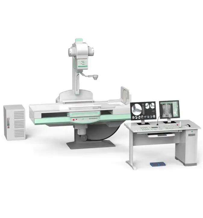
X-Ray Windsong utilizes the newest digital X-Ray systems that will expose both adult pediatric patients to a significantly reduced level of radiation yet with amazing images and clinical capabilities such as. They provide images of the inside of the body in still form.
 What Is The Difference Between Fluoroscopy And Radiography
Explain the working principles of the image.

X ray fluoroscopy. X-rays are static images. It is based on an x-ray image intensifier coupled to a stillvideo camera. Start Your Certification Today.
X-Ray fluoroscopy XRF determines bulk composition using x-rays to stimulate characteristic x-ray emission. Patients screen for prolonged time requires substantial reduction in X-ray exposure. Risksbenefits of fluoroscopyBecause Fluoroscopy is an x-ray machine it has the same risks as other x-ray machines.
Today fluoroscopy is used foAcquisition parameters referencer diagnosis and guidance of clinical procedures. Denoising is essential to enhance fluoroscopic image quality and can be considerably improved by considering the peculiar noise characteristics. Philips x-ray fluoroscopy solutions are highly customizable.
Fluoroscopy is an innovative technology that offers many benefits using X-rays. Fluoroscopy is a technique used to capture X-ray images in motion. The plate is connected to an image.
Two major risksThere is a small possibility of developing cancer due to the exposure to the radiationInjuries such as burns caused by the radiationBenefitIf a patient is in need of a Fluoroscopy the benefit outweighs the minute risks. Start Your Certification Today. The limited number of photons generates relevant quantum noise.
Fluoroscopy is a study of moving body structures--similar to an X-ray movie A continuous X-ray beam is passed through the body part being examined. Radiography and Fluoroscopy Zhihua Qi PhD LEARNING OBJECTIVES 1. The beam is transmitted to a TV-like monitor so that the body part and its motion can be seen in detail.
From portable x-ray equipment to complete digital X-ray rooms we can provide a solution that fits your workflow and budget. In XRF the maximum incoming x-ray energy varies with manufacturer and instrument configuration but typically ranges from 50 to 60 kV. Ad Find Local and Online Radiology Schools Fast.
Shortly after the discovery of X-rays in the late nineteenth century fluoroscopy was developed to enable visualization of moving anatomy. They both are powered by electromagnetic radiation for the purpose of obtaining necessary images. Applications of fluoroscopy are found throughout medicine including radiology cardiology urology.
Describe the functions of the main components of an X-ray tube including the cathode the anode the collimator and the filters. It creates the images in a video form which can open many other opportunities for its usefulness. Fluoroscopy is an imaging modality that allows real-time x-ray viewing of a patient with high temporal resolution.
At Philips our X-ray and Fluoroscopy equipment offer excellent workflow and quality images to drive through-put and confident diagnoses while enabling high staff and patient satisfaction. A portion of the x-rays are absorbed or scattered by the internal structure and the remaining x-ray pattern is transmitted to a detector so that an image may be recorded for later evaluation - UCLA Dept of Radiology. It uses the same technology as an X-ray to generate a working image for a doctor to interpret and decide a course of treatment for the patient.
Radiography Fluoroscopy Systems. Theres no limit to what we can do together. Describe the roles of an X-ray generator and its desired characteristics for medical imaging.
X-ray and fluoroscopy technology are essentially the same with a few notable differences. Ad Find Local and Online Radiology Schools Fast. During a radiographic procedure an x-ray beam is passed through the body.
During a fluoroscopic procedure a radiologist transmits a low energy X-ray beam lower energy than traditional X-rays through the patient onto a plate or carriage. Connecting data technology and people. In recent years flat panel detectors which are similar.
Subtraction of an image with bones for an unobstructed view of soft tissue and seamless long bone or spine images with one automated exam that features auto image paste.
